


Imaging diagnosis in germ cell and sex cord-stromal tumors
Norbert Stachowicz, Dorota Morawska, Jan Kotarski
 Affiliacja i adres do korespondencji
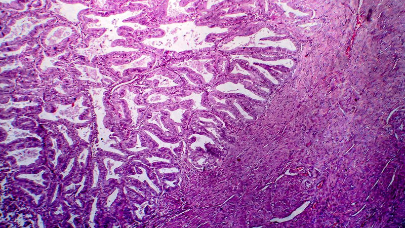
Clinical diagnosis of ovarian tumors is the more difficult, the earlier is the clinical stage at presentation. On the other hand, while early-stage conditions are much more difficult to detect, early diagnosis is associated with the best outcomes. Rare sex cord-stromal and germ cell tumors present sonographic features similar to other ovarian tumors. Sonographic report should address the following: location, size and echogenicity of ovaries, location of adnexal lesions and their relation to other pelvic structures, precise description of solid elements, septations and papillomatous excrescences within the tumor as well as presence of free fluid within the peritoneal cavity. In suspected malignant tumors, most useful prognostic features include papilliform excrescences, so their location, size and number must be always documented. Furthermore, number and thickness of septations is of great prognostic significance. Description of tumor morphology should include its external and internal walls. Sonographic diagnosis may predict malignancy of lesion based on such features as solid areas, papilliform excrescences, septations or abnormal pattern of tumor vasculature. Due to neoangiogenesis in malignant tumors, study of vasculature and perfusion is also important. A typical finding in a malignant tumor is increased late-diastolic flow velocity, reduced resistance index RI to less than 0.5 and pulsation index to less than 0.8. Frequent is also elevation of peak systolic velocity PSV to over 12 cm/s. In spite of sophisticated and continuously improved imaging techniques in sonography, including “color” and “power” options in angio-Doppler studies as well as introduction of 3D/4D technique, malignant process may at best be suspected. Final diagnosis of malignant tumor is always based on histological studies.
